TPLO (Tibial Plateau Leveling Osteotomy)
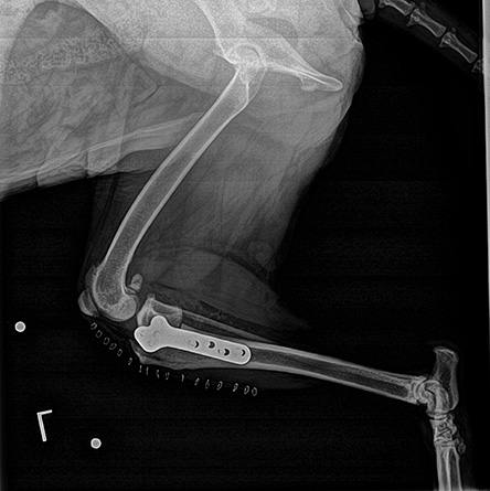
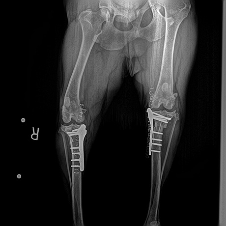
This surgery changes the angle and relationship of the femur and the tibia. The overall intent of the surgery is to reduce the amount that shifts forward during a stride. This is accomplished by making a semicircular cut through the top of the tibia. The top section of the tibia is then rotated backward until the angle between the tibia and femur is deemed “appropriately level,” A metal bone plate is then used to affix the two sections of tibia in the desired positions, allowing the tibia to heal in its new configuration.
This realignment of the surfaces within the stifle helps to provide stability during a stride, and helps to reduce future joint inflammation and OA. By carefully adjusting the angle or slope of the top of the tibia, surgeons are able to replicate a more normal configuration of the knee joint and reduce mechanical stress.
TPLO surgery “levels” the tibia to prevent the femur from sliding backward, thereby stabilizing the joint. Pictured below is an example of a left leg TPLO and then a bilateral (both legs) TPLO.


Tightrope Technique
The newest procedure for repairing a ruptured CCL by ligament replacement is the TightRope Ligament technique. This procedure has been shown to be highly effective. The TightRope CCL technique is minimally invasive, and more cost effective in comparison to the TPLO or TTA. The data suggest that TightRope can be successfully performed in medium, large and giant breed dogs resulting in outcomes which are comparable to TPLO or TTA.
The TightRope CCL counteracts the forward tibial thrust and inward rotation resulting from CCL damage, while providing optimal joint range of motion. This procedure mimics the natural cruciate ligament functions, perhaps better than any other procedure developed to date.
The artificial TightRope Ligament is an ultra-high strength, flat, smooth, braided, ribbon-like ligament composed of a multi-stranded long chain ultra-high molecular weight polyethylene core with a braided jacket of polyester giving it unsurpassed strength, virtually eliminating ligament breakage. This procedure provides clients another option when a less invasive, less radical and somewhat more cost effective procedure that does not cut bone is desired by the pet owner.
Extracapsular Technique
The knee joint is first inspected via a small incision (arthrotomy) or insertion of a camera (arthroscopy). The torn remnants of the ACL are removed and the meniscal (diagram) structures are examined. If the meniscus is torn, the damaged portion is removed.
A strong, non-absorbable suture (nylon band) is passed around a small bone on the back of the knee and then placed through a small hole made in the front of the tibia bone. The suture is then tightened which serves to prevent drawer motion, effectively taking over the job of the torn cruciate ligament.
Over time, scar tissue will form around the suture which helps to stabilize the joint. This technique is most commonly used in small dogs and cats. About 85% of patients show a significant improvement after surgery and are able to resume pre-injury activities.
Tibial Tuberosity Advancement (or TTA)
The TTA, or tibial tuberosity advancement, is explanatory also by its name. Tibial = relating to the tibia, the lower half of the of the stifle joint. The tuberosity is the front edge of the tibia just below the stifle joint where the patellar tendon attaches, and advancement, meaning to bring forward. The tibial tubersosity (patellar tendon attachment) is just moved forward a certain amount and a spacer is screwed in place to hold it together until the bone cut is healed. The amount of advancement is determined by a right angle 90o of the patellar tendon to the tibial plateau. By advancing the tibial tuberosity to a 90o angle, the stifle joint is stabilized by shifting the weight to the caudal cruciate ligament. In effect there is no longer a need for the cranial cruciate ligament.
Appointments
Our appointment book is computerized which allows us to efficiently make appointments for you and your pet. Our receptionists and team will attempt to accommodate all requests to the best of our ability. Emergencies are accepted anytime our clinic is open. Emergencies can be things such a snail bait poisoning, hit by car, and chocolate ingestion. If you feel you have an emergency with your pet, it is best to call before coming in so that a staff member can advise you on your particular emergency.
We are also available for urgent care when the condition is not life-threatening, but you feel your pet needs to be seen before you are able to get an appointment. Our veterinarians will work to “squeeze” you in between scheduled appointments. When you arrive, our receptionists will be able to give you an estimate on how long you may have to wait in order to be seen.
Payments
Edna Valley Veterinary Clinic and Surgical Services accepts payment via cash, check, MasterCard, Visa and Care Credit. So that we can continue to provide you high quality service utilizing the best medical technologies, we request that payment be made at the time services are rendered.
We also recommend looking into pet insurance for your pet. A good place to start is here:
Discounts
Edna Valley Veterinary Clinic and Surgical Services offers a free exam for pets adopted from San Luis Obispo Animal Services or Woods Humane Society. Pre-approval is required to qualify for the rescue group discounts, please call us for more information.

